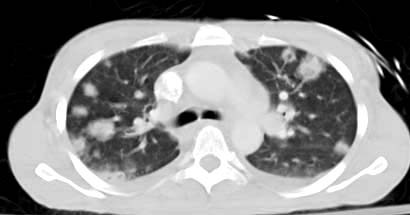
Septic pulmonary emboli is embolization of intravascular
thrombus containing microorganisms into the lungs. Septic emboli can occur from
varying sources like tricuspid valve
endocarditis, infection elsewhere in the body with
associated septal defect, infected deep venous thrombosis, venous lines /
central venous catheters, pacemaker wires etc.
On
CT chest, "vessel
sign" is interesting to watch with peripheral
nodules with clearly identifiable feeding vessels. May be visible are -
subpleural nodular lesions or wedge-shaped densities with or without necrosis
caused by septic infarcts.
No comments:
Post a Comment